
Glaucoma
Glaucoma is a condition that can cause progressive damage to the optic nerve, often due to increased pressure inside the eye, and can lead to vision loss if left untreated.
What is glaucoma?
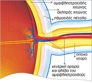
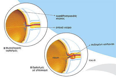
Glaucoma is a heterogenic group of diseases that are characterized by an irreversible insult to the optic nerve. The optic nerve consists of retinal nerve fibers that carry the optical information from the eye to the brain (Fig. 1).
Glaucoma is usually asymptomatic and typically the glaucoma patient is unaware of his visual loss until late stages of the disease. The reasons of unawareness are that interval changes occur at a slow rate on a daily basis and that patients develop coping strategies to fill in their peripheral visual field loss by “scanning” the environment.
For this reason, a preventive evaluation by a glaucoma specialist is essential in confirming the diagnosis early in order to prevent further damage, maintain vision and vision related quality of life. Therefore, the American Academy of Ophthalmology (AAO) recommends annual screening for people over forty years of age, especially for those who have a family history of glaucoma.
The Etiology of Glaucoma
The etiology of glaucoma is unknown. However, in most cases the optic nerve is damaged as a result of elevated intraocular pressure. Reduction of patient’s intraocular pressure by medications, laser or surgery is of paramount importance in our treatment strategy.
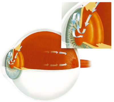
Αqueous humor (fluid within the eye) is constantly produced and drained. The balance between production and outflow is reflected by the intraocular pressure (IOP). If for some reason outflow is obstructed there is an increase in intraocular pressure (Fig. 2). Patients with elevated IOP without any evidence of structural or functional damage to the optic nerve suffer from ocular hypertension a significant risk factor for the development of glaucoma. Concurrent damage to the optic nerve establishes the diagnosis of glaucoma. However, not all patients with ocular hypertension will develop glaucoma vice versa some patients will develop glaucoma in the absence of elevated IOP (low tension glaucoma).
There are several other epidemiologic risk factors predisposing to glaucoma development such as advanced age, family history of glaucoma, race, use of oral contraceptives or corticosteroids, the presence of vascular diseases etc.
Recently, multicenter clinical trials (i.e. Ocular Hypertension Treatment Study) have demonstrated the importance of a new risk factor: the thickness of the central cornea. Patients with a thinner cornea carry an increased risk of developing glaucoma. At the Athens Vision Eye Institute, we employ contact and non-contact methods to precisely measure central corneal thickness in all patients with glaucoma and ocular hypertension.
Glaucoma: Diagnosis, Evaluation and Management
Diagnosis of glaucoma does not rely on measurement of the intraocular pressure alone. Elevated eye pressure (>21mmHg) does not automatically imply glaucoma. On the other hand, normal intraocular pressure (<21mmHg) does not preclude the diagnosis of glaucoma. In fact, 1/3 of Caucasian patients with glaucoma do not ever demonstrate untreated eye pressures above 21mmHg.
Diagnosis of glaucoma
Patients with glaucoma demonstrate a characteristic optic nerve head appearance and evidence of progressive loss over time. Therefore, the diagnosis of glaucoma is based on a comprehensive eye examination that includes individual medical and ocular history, family history and ancillary testing, the most pertinent of which are listed below:
- Tonometry
- Gonioscopy
- Stereoscopic evaluation of the optic nerve head and retinal nerve
fiber layer. - Visual field testing.
Tonometry
Accurate measurement of the intraocular pressure is of vital importance in glaucoma management.
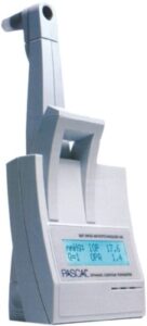
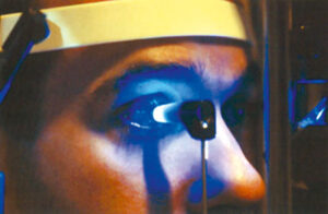
At the Athens Vision Eye Institute in addition to the widely used Goldman applanation tonometer we use three additional new devices: the digital Pascal tonometer which is minimally affected by corneal elastic properties and is therefore very useful in patients who have undergone laser refractive surgery, the non-contact Ocular Response Analyzer (ORA) that offers additional information on the ocular viscoelastic properties and the eye-Care tonometer that does not require eyedrops and is therefore very useful in children.
All three tonometers provide additional reliable information about the eye pressure while minimizing the impact of factors that vary from patient to patient like corneal thickness and corneal resistance.
Evaluation of the Optic Nerve and Retinal Nerve Fiber Layer
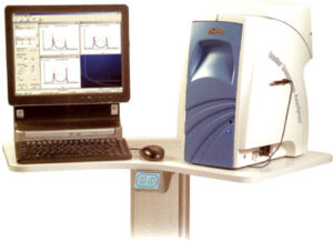
Objective and quantitative methods of evaluating the optic nerve head anatomy have emerged recently such Optical Coherence Tomography (OCT), Scanning Laser Polarimetry (GDx), Confocal Scanning Laser Ophthalmoscopy (HRT). Currently, we can precisely image the morphology of the optic nerve head, measure the thickness of the retinal nerve fiber layer and obtain useful information for the diagnosis of glaucoma. There are normative databases allowing comparisons with normal aged-matched individuals and the instruments can be used for follow-up of established patients over time.
At the Athens Vision Eye Institute, we operate three of the latest generation OCTs (Fourier domain OCTs) with a resolution of 4-5 μm, high processing speed and the ability to three-dimensionally reconstruct the optic nerve head.
It is also possible to appraise the retinal ganglion cell layer in the macular region obtaining useful information regarding the state of the disease and to quantify the vascular supply to the optic nerve head non-invasively.
The precise documentation of these anatomical features contributes to the early diagnosis of glaucoma, since structural optic nerve changes precede functional loss in glaucoma.
Visual Field Testing (Standard Automated Perimetry)
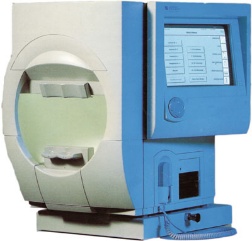
The peripheral visual field performance is predominately affected in early and moderate glaucoma before patients become symptomatic. Visual field testing (perimetry) is of paramount importance in glaucoma detection and follow-up. At the Athens Vision Eye Institute, we use the latest edition of the Humphrey Visual Field Analyzer, which has been employed in most multicenter glaucoma clinical trials worldwide. Up-grated software (Glaucoma Progression Analysis and Visual Field Index) permits early diagnosis of glaucoma and state of the art follow-up utilizing sophisticated statistical analysis.
By abiding to European and American Glaucoma Society recommendations and high-quality standards we offer evidence based clinical management to the benefit of our patients.
Ask our physicians how based on a given number of visual field tests they can infer reliable predictions about visual performance in the future.
Well-informed patients do not only inquire about their intraocular pressure. They are similarly concerned about the optic nerve and the visual fields!
Glaucoma: Treatment
Medical Treatment for Glaucoma
Glaucoma Laser Treatment
Surgical Treatment for Glaucoma
- Glaucoma affects the optic nerve, the cable that connects the eye to the brain. Glaucoma is in its early stages asymptomatic and cannot be appreciated by the patient. Timely diagnosis relies on appropriate screening examinations.
- The destruction of optic nerve in glaucoma is irreversible.
- Elevated intraocular pressure is a major contributing factor but not the only cause of glaucoma.
- Appropriate glaucoma management involves regular systematic monitoring of the optic nerve along with visual field testing with concomitant intraocular pressure measurements. The examination intervals vary upon type and stage of disease and patients’ demographic characteristics.
Doctors who specialize in this condition

Gerasimos Kopsinis