Keratoconus
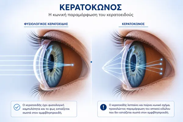
Keratoconus is an eye condition that affects the shape—or more precisely, the curvature—of the cornea, the transparent front surface of the eye that helps focus incoming light.
What is keratoconus?
Keratoconus is an eye condition that affects the shape—or more precisely, the curvature—of the cornea, the transparent front surface of the eye that helps focus incoming light.
Causes of Keratoconus
Symptoms of Keratoconus
- Blurred vision
- Progressive myopia and astigmatism
-
In the early stages, glasses may provide satisfactory vision, but as the condition progresses, they may no longer provide adequate visual correction.
Diagnosis of Keratoconus
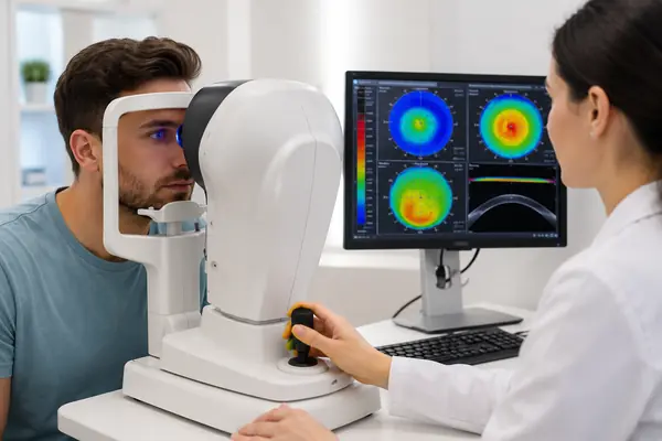
Corneal Topography / Corneal Wavefront (CORWAVE®)
Treatment
Glasses
Contact Lenses
- Soft lenses: limited effectiveness
- Rigid gas-permeable (RGP) lenses: better vision and comfort, suitable for long-term visual correction
- Hybrid lenses: combine the comfort of a soft lens with the visual quality of a rigid lens
Surgical Treatment
-
UVA Cross-Linking (collagen cross-linking): the only treatment specifically designed to halt or slow the progression of keratoconus by strengthening the biomechanical stability of the cornea. It is performed under topical anaesthesia (eye drops) and is generally well tolerated. With modern accelerated protocols, the procedure takes about 30 minutes.
-
Astigmatic intraocular lenses: these are implanted inside the eye to improve vision in selected cases.
-
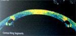
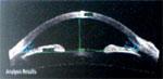
Intrastromal corneal rings: small implants that modify corneal curvature and improve vision in selected patients, with effects that may be maintained over the long term.
-
CAIRS (allogeneic corneal rings): implants made from donor human corneal tissue, offering excellent biocompatibility, and placed using a femtosecond laser.
-
Keratoplasty (corneal transplant): replaces the affected corneal tissue with a donor graft. Keratoplasty may be full-thickness (penetrating keratoplasty) or partial-thickness (anterior lamellar keratoplasty).
Frequently Asked Questions About Crosslinking
Collagen cross-linking (Collagen Cross-Linking with UVA–Riboflavin) is currently the only treatment specifically designed to increase the biomechanical stability of the cornea without the implantation of donor tissue (e.g., partial-thickness or penetrating keratoplasty) or synthetic implants (e.g., intrastromal corneal rings). This strengthening is achieved by creating additional cross-links between collagen fibres through controlled exposure to ultraviolet A (UVA) radiation. Riboflavin acts as a photosensitizer, facilitating the cross-linking reaction while helping to limit UVA exposure to deeper ocular structures.
This method of strengthening the corneal tissue is recommended for individuals with keratoconus, other forms of corneal ectasia, and for stabilizing corneal curvature in cases of corneal instability following excimer laser refractive surgery. It also may have a role in severe corneal infections and inflammatory conditions, although its use in such cases is still considered investigational.
The primary goal of the treatment is to stabilize corneal shape and curvature, that is, to halt the progression of keratoconus or corneal ectasia. Thus, eyes with satisfactory vision prior to treatment can achieve increased biomechanical stability, significantly reducing the risk of further disease progression. In eyes with moderate keratoconus, a statistically significant reduction in myopia and astigmatism may be observed on corneal topography, averaging about two diopters, often developing gradually over several months following treatment. In cases of advanced keratoconus, however, collagen cross-linking alone is usually not sufficient to provide satisfactory visual improvement. In selected cases, collagen cross-linking is performed simultaneously with or several months before selective surface photokeratectomy, with the aim of improving vision and avoiding keratoplasty, at least temporarily.
Corneal collagen cross-linking in its current form resulted from the pioneering research of Theo Seiler’s team and collaborators at the University of Dresden in the 1990s (leading to the “Dresden protocol”). The method received CE marking in Europe in 2006, and in 2016 it was officially approved by the U.S. FDA (KXL System with Photrexa solution). Today, after more than two decades of clinical use and hundreds of thousands of treatments worldwide, collagen cross-linking is an established first-line treatment for progressive keratoconus, with multiple CE-marked devices available and several specialized protocols (accelerated, transepithelial, PACK-CXL for infections, as well as protocols for thin corneas).
A decrease in visual acuity in eyes with keratoconus, an increase in astigmatism and myopia or changes in keratometric or topographic indices, as well as a reduction in corneal thickness, usually provide sufficient evidence that the cornea is unstable and the disease is progressing. In these cases, a well-informed patient may choose to undergo corneal strengthening with collagen cross-linking.
The procedure is performed under topical anaesthesia using eye drops and is generally well tolerated during treatment. The ophthalmologist removes the epithelium from the central cornea (epi-off technique) to allow riboflavin to penetrate the corneal stroma. Riboflavin drops are then instilled for approximately 10 minutes, corneal thickness and adequate riboflavin saturation are assessed, and the cornea is then exposed to UVA radiation (370 nm). The duration of irradiation depends on the protocol used (classic Dresden protocol: 30 minutes; accelerated protocols: 5–10 minutes). Throughout the procedure, the cornea is hydrated with riboflavin drops and saline solution, and after the treatment is complete, a bandage contact lens is placed and remains in place for 3–5 days until re-epithelialization.
The application of UVA radiation may, at least theoretically, have a harmful effect on the corneal endothelium, particularly if established safety parameters are not respected. Although the corneal epithelium usually heals within 2–5 days, its removal may cause discomfort, a foreign body sensation, burning, as well as tearing, redness, and blurred vision. Although these symptoms are usually temporary and expected, the possibility of corneal infection (infectious keratitis), although rare, should always be considered. When established treatment protocols and safety thresholds are followed, UVA exposure to deeper ocular structures is minimized, and complications such as lens damage or cataract formation are not expected.
Although several centers have already begun performing bilateral treatment on the same day, at Athens Vision we recommend treating the more severely affected eye first, provided treatment is indicated and all safety criteria are met. The other eye can undergo the same treatment a few weeks later. As for the patient’s age, this is not in itself a determining factor. We have no reason to postpone treatment in a 14-year-old patient when there is clear evidence of progression in corneal measurements, particularly as such changes may be irreversible.
Available follow-up data indicate long-term stability and improvement in keratometric indices for many years after a single treatment. Depending on the severity of the condition, age, and the patient’s response to the initial treatment—and given the natural remodelling of corneal collagen—we believe that very young patients may require repeat treatment three to ten years after the initial procedure.
Recent developments in collagen cross-linking
2. Astigmatic Intraocular Lenses
Their use is indicated in cases of non-progressive keratoconus where vision with very high degrees of myopia/astigmatism is satisfactory. These lenses are usually placed in the anterior chamber of the eye through a small incision using microsurgery, and the visual result is immediate when the patient has been properly selected. Furthermore, as we will see below, the use of such lenses in combination with other techniques has helped patients achieve very good results without a transplant in a much shorter period of time.
3. Intrastromal Corneal Ring Segments
Advantages
Disadvantages
4. CAIRS — Allogeneic Intrastromal Corneal Rings
5. Keratoplasty
a. Penetrating keratoplasty
Partial-thickness keratoplasty
In the case of keratoconus, the diseased anterior corneal stroma is removed, while the patient’s Descemet membrane and corneal endothelium are preserved. After the cornea is dissected to the required depth using specialised microsurgical instruments, automated microkeratomes, or selected laser systems, the surgeon removes the anterior diseased corneal lamella and replaces it with a donor anterior lamellar graft from which Descemet membrane and the endothelium have been removed.
Frequently Asked Questions
In a corneal “transplant,” whether full-thickness or partial-thickness, a portion of the corneal tissue is replaced. Therefore, it is a tissue transplant rather than an organ transplant, such as a kidney transplant. Corneal transplantation is generally performed to restore or improve vision and quality of life rather than as a life-saving procedure.
Consequently, graft failure does not pose a threat to the patient’s life, and in most cases a failed graft can be treated or replaced with another corneal graft. If the procedure fails, repeat transplantation is usually possible.
In cases of partial-thickness corneal transplantation, the risk of rejection is significantly reduced; in anterior lamellar keratoplasty, endothelial rejection is eliminated because the patient’s own endothelium is preserved. Furthermore, because the procedure does not enter the anterior chamber, the patient also benefits from other important advantages, which are discussed below.
In the case of penetrating or partial keratoplasty, the procedure is performed using a graft from the eyes of recently deceased donors. The European Union and the U.S. have defined in detail the process of donor tissue selection, testing, and transport to the surgical centre where the procedure will take place, as well as the monitoring of the long-term outcome of the procedure by non-profit organizations known as “eye banks.” Accredited eye banks operate under strict supervision and clearly defined operating standards and regulations (similar to blood banks) to ensure that the screening of tissue grafts destined for our patients’ eyes is always thorough and comprehensive. Athens Vision works exclusively with eye banks approved by the relevant authorities in the European Union and the United States.
Unlike organ transplants, where the coordination of all parties involved in the procedure is particularly important, time is less critical in corneal transplantation. Corneal grafts can be retrieved several hours after the donor’s death, may remain at the eye bank for testing for several weeks when in vitro culture methods are used, and can be transported by air on ice from the eye bank to surgical centres worldwide.
As a result, in the U.S. alone, over 35,000 transplants are performed annually, and there is a surplus of more than 10,000 grafts each year that can be exported to other countries. Very careful studies have shown that grafts transported by air show no disadvantage in graft survival compared with grafts implanted at transplant centres located close to an eye bank.
Grafts are rejected relatively rarely. In the case of keratoconus, the probability of rejection is less than 5%. The reason rejection is relatively uncommon is that corneal grafts lack blood vessels and are sutured onto tissue that is also avascular. For this reason, in routine corneal transplantation, no tissue compatibility testing is performed, nor are donor and recipient blood groups routinely matched.
Postoperative treatment consists of topical eye drops every few hours during the first few days. Treatment is usually discontinued after 3–4 months. Only in a few exceptional cases is additional preoperative or postoperative treatment required.
The complications of keratoplasty (apart from graft rejection) are the same as those of any other intraocular surgery, such as cataract surgery. These include infection, inflammation, bleeding, increased intraocular pressure, and complications involving the posterior segment, such as suprachoroidal haemorrhage and retinal detachment. These are extremely rare, and even when they occur, they are usually treatable. Because in partial-thickness keratoplasty the procedure remains extraocular, without entering the anterior chamber, and involves only the cornea, these complications are not typically associated with partial-thickness keratoplasty.
However, in addition to these complications, special mention should be made of the development of postoperative astigmatism. In cases of keratoconus transplantation, postoperative astigmatism is particularly common and occurs in the majority of keratoplasty procedures (average 3.5 D).
Surgical experience, preoperative measurements, the patient’s age, the size of the graft, the cutting instruments, and intraoperative topography—no matter how important they may be—do not guarantee clear vision without glasses or contact lenses in every case.
The patient returns home the same day, and the protective bandage is removed during the first postoperative examination the following day. Although visual acuity improves gradually over several months after surgery, the patient does not need to remain in bed after the procedure. Depending on the level of preoperative vision in the treated eye—which is usually severely impaired—as well as the visual function of the fellow eye, the patient can return to work relatively quickly, provided that the necessary protective measures are taken, including protective eyewear.
As for anterior lamellar keratoplasty, the advantages of the technique include the avoidance of intraocular manipulation during the procedure, which in practice means no risk of endothelial rejection and a much lower likelihood of endophthalmitis. Additionally, in the case of partial transplantation, the sutures remain in place for several months fewer, resulting in less irritation and a lower risk of infection. In this case, the need for corticosteroid eye drops (immunosuppression) is dramatically reduced, thereby reducing the risk of corticosteroid-induced cataracts and increased intraocular pressure, while further reducing the risk of infection. Anterior lamellar keratoplasty is also superior in the event of future ocular trauma, since the strength of the cornea more rapidly approaches that of a normal cornea—something that is never the case with conventional transplantation.
Equally important for both the patient and the surgeon is the ability to use grafts from older donors, with no limitations related to endothelial cell survival, simply because the donor’s endothelium is not transplanted to the patient. Especially in keratoconus, a lamellar transplant from an adult donor is preferred because the cornea, by definition, has more compact and cross-linked collagen and therefore—at least theoretically—may provide better results. In conventional transplantation, although we always avoid grafts from very young donors in cases of keratoconus, we prefer grafts from donors in their 30s or 40s given the typically young age of patients with keratoconus.
The disadvantages of partial or lamellar keratoplasty are the steep learning curve of the technique and the fact that, in 5–10% of cases, the procedure must be interrupted or converted to a conventional transplant if an intraoperative rupture of Descemet’s membrane is detected. Furthermore, conventional transplantation does not appear to be inferior to partial transplantation in terms of postoperative vision.
In both cases, it is possible to use customised excimer laser surface photoablation using data from the corneal surface wavefront (CORWAVE) to achieve better results. However, it appears that because the biomechanical stability of the cornea is greater with a partial transplant, the long-term results are likely to be better after partial keratoplasty when selective surface photoablation is also performed.
In older grafts, after examining endothelial status, corneal thickness, and the repeatability of keratometric measurements, it is often possible to further improve vision using customised excimer laser surface photoablation with data from the corneal surface wavefront (CORWAVE). Furthermore, significant visual improvement may also be achieved with toric intraocular lenses in selected cases. Re-transplantation is also a viable solution but is rarely our first choice.
A doctor who specializes in this condition