
SERVICES / CORNEA

Cornea

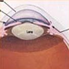
The cornea is the transparent front layer of the eye.
What is the cornea?

The cornea is the transparent front layer of the eye.

Although it is the most densely innervated tissue in the eye (and is therefore highly sensitive), it normally lacks blood vessels. Due to its curvature and transparency, it works in conjunction with the eye’s lens—which makes a significant but smaller contribution—to focus light onto the photoreceptors of the retina.

The basic structure of the cornea is simple.

It consists of:

  • the epithelium

  • the stroma,

  • the endothelium.

Corneal opacity, or a significant change or abnormality in its curvature (e.g., keratoconus), has a direct impact on visual quality. Corneal transplantation is often the best treatment option, because many inflammatory or non-inflammatory conditions, injuries, and infections leave permanent scarring and corneal opacity.

What is a corneal transplant?

A corneal transplant (or keratoplasty) is the replacement of all or part of the cornea. Depending on the corneal layer being replaced, the procedure is classified as partial-thickness (anterior or posterior lamellar keratoplasty) or full-thickness (penetrating keratoplasty). If a synthetic implant is used, the procedure is called keratoprosthesis (artificial cornea implantation).

Penetrating keratoplasty (PKP)

Corneal transplantation is the most successful human tissue transplant. It is performed at least four times more often than kidney transplantation and has the highest success rates (up to 95% in certain conditions). In the vast majority of cases, no postoperative oral medication is required (topical eye drops are usually sufficient), and most often the procedure is performed under local anesthesia alone; after the operation, the patient returns home the same day (outpatient surgery).

The procedure to remove the central part of the cornea is performed using specialized corneal trephines, usually after the administration of local anesthesia. The procedure continues with additional intraocular procedures, if necessary, such as cataract extraction or iridoplasty, and is completed by suturing the graft to the recipient’s cornea using extremely fine microsurgical sutures, thinner than a human hair.

Partial-thickness keratoplasty

Where possible, we prefer selective partial-thickness replacement of the cornea. It is possible to selectively replace the anterior corneal layers, nearly the full corneal thickness, or only the endothelial layer. The separation is performed using automated microkeratomes and specialized microsurgical instruments.

Anterior lamellar keratoplasty

(DALK, ALTK)
Only the anterior layers of the cornea are selectively replaced. The graft acts like a biological contact lens that is surgically placed onto the cornea rather than within the eye. The first near-full-thickness anterior lamellar transplant was performed by Anastasios K. Charonis at the Orasis Center in September 2002.
Regarding anterior lamellar keratoplasty, the advantages of the technique can be summarized as follows: avoidance of intraocular procedures, no risk of endothelial rejection, the possibility of leaving sutures in place for a longer period, no need for immunosuppressive medication, and significantly greater corneal structural integrity in the event of future trauma. At Athens Vision (Dr. Anast. K. Charonis), the first anterior lamellar keratoplasty in Europe using an excimer laser with the SCHWIND AMARIS system was performed in 2008. This method allows for excellent dissection of the posterior stromal lamella when the Big Bubble technique cannot be used. At Athens Vision, we have 15 years of experience using customized excimer laser photoablation on the graft for even better results.
This method is ideal for the surgical treatment of relatively advanced keratoconus, which typically affects young patients who may be at risk of ocular trauma in the future.

New developments in anterior lamellar keratoplasty

At Athens Vision, we believe that the future of keratoconus treatment lies in minimally invasive microkeratoplasty, that is, the fabrication of a micro-graft approximately as thin as a conventional contact lens that will be epithelialized—meaning it will sit on the patient’s corneal stroma beneath the epithelium, without sutures. (Athens Vision Minimal Additive Refractive Treatment—AMART) A specific method for calculating and fabricating in vivo this biological contact lens has been patented by us, and in 2021 it was licensed/transferred for further evaluation and commercialization to a major refractive laser manufacturer.

Posterior lamellar keratoplasty / Endothelial keratoplasty

(DSEK, DSAEK, DMEK)
In the case of selective replacement of the endothelial layer, the procedure takes just 20 minutes, no sutures are required, postoperative astigmatism is negligible, and visual recovery is much faster than with conventional transplantation. Epithelial defects and suture-related complications are therefore avoided. The first posterior lamellar keratoplasty in Greece was performed in December 2002 by Anastasios K. Charonis. Twenty years later, endothelial keratoplasty has become one of the most widely performed forms of keratoplasty in Europe and throughout the developed world.
In the DSEK procedure, the surgeon or the tissue bank prepares the donor cornea by creating a thin posterior lamellar graft using microsurgical instruments, an automated microkeratome, or, in selected techniques, a femtosecond laser. The partial-thickness graft is placed by the surgeon in its final position within the anterior chamber, behind the cornea, after the diseased portion of the recipient cornea (Descemet’s membrane and endothelium) has been removed. No sutures are used to secure the graft, which is held in place by an air bubble. Disadvantages of the technique include intraoperative endothelial cell loss and possible graft detachment or displacement during the first few postoperative days. In case of failure, it is possible to perform a new partial-thickness or, more commonly, full-thickness keratoplasty. Air/gas tamponade and early graft detachment are well-recognized features of endothelial keratoplasty.
A variation of the method involves transplanting only Descemet’s membrane together with the endothelial cells attached to it. This is the DMEK procedure (Descemet Membrane Endothelial Keratoplasty). Since 2017, DMEK has been the most common transplant procedure recommended to Athens Vision patients with endothelial dysfunction, such as Fuchs’ dystrophy or bullous keratopathy. DMEK has an advantage over DSEK/DSAEK because it is associated with lower rates of graft rejection. The DMEK graft, with a thickness of approximately 10–20 microns, is among the thinnest grafts transplanted in modern medicine. DMEK grafts consist of Descemet membrane and endothelium without donor stroma, typically measuring around 10–15 μm.

The DSO/DSO Xtra procedure

The first DSO (Descemet Stripping Only) procedure was performed in Greece in 2011 by Anastasios K. Charonis at the Athens Vision Day Surgery Unit.

During this procedure, the central diseased Descemet’s membrane and endothelium are removed without replacing them with a graft. Today, after ten years of experience at Athens Vision, we know that this method should be attempted only in selected cases that meet specific criteria associated with a higher likelihood of success. For example, it is indicated for well-informed patients with Fuchs’ dystrophy classified as Krachmer stage III–IV, with an adequate peripheral endothelial cell count and the option of treatment with ripasudil (DSO Xtra).

Surgery with alloplastic adsorption barriers (Endoart Keratoplasty)

Surgery using alloplastic adsorption barriers is an experimental but groundbreaking approach to the problem of corneal endothelial layer insufficiency, currently being investigated by Athens Vision and the Institute of Research and Technology of the (ITE-FORTH) on experimental rabbits. Our in vivo protocol was conducted at the N. S. Christas Laboratory of Experimental Surgery and Surgical Research with spectacular results between April 2021 and February 2022. The project was partially funded by the General Secretariat for Research and Technology (GSRT) / NSRF 2014-2020. This procedure has not yet been performed on humans but is expected to provide at least a temporary, functional, and cost-effective solution to the problem of corneal blindness until a suitable human graft is found. Project Coordinator: Anastasios K. Charonis. In 2024/25, the first four surgeries using alloplastic implants (instead of the classic endothelial keratoplasty—DSEK/DMEK/PDEK) were performed with complete success in Greece at the AthensVision Medical Institute of Ophthalmology.

Keratoprosthesis: the artificial cornea

The artificial cornea is a laboratory-manufactured artificial corneal implant designed to replace the diseased central portion of the cornea and restore vision. This artificial implant is surgically implanted into the patient’s eye using microsurgical techniques that closely resemble a standard corneal transplant procedure.
To date, rigid materials are preferred because of their superior clinical performance and outcomes compared with soft-material corneal prostheses.

When is the use of an artificial cornea preferred?

Typically, keratoprosthesis implantation is reserved for patients with severe vision loss due to advanced corneal disease. The patient must be fully informed about both the advantages and the potential risks and limitations of the procedure. The decision to proceed with surgery must always be individualized. In general, keratoprosthesis implantation may be indicated in the following cases:

What are the advantages of using a keratoprosthesis?

What are the disadvantages of using a corneal prosthesis?

The keratoprosthesis we currently use at Athens Vision (Boston Keratoprosthesis I & II) has some of the best documented outcomes in the international ophthalmic literature, backed by 35 years of international experience and continuous improvements. With careful patient selection, counselling, and follow-up, keratoprosthesis implantation is a realistic option for substantial visual improvement and, in selected cases, restoration of useful vision.

Research and Innovation at Athens Vision

Beyond our daily clinical practice, at Athens Vision, we maintain an active research and innovation portfolio in collaboration with Greek and international academic institutions and technology partners. The areas we focus on include refractive and corneal surgery with computationally guided personalization, advanced premium intraocular lens solutions for cataract surgery, non-invasive monitoring of ocular parameters using photonic technologies, and the application of cold atmospheric plasma in ophthalmology.
Some of these projects are currently undergoing patent protection or involve confidential collaborations with industry partners and, for this reason, are not presented publicly in detail. As in the past—a prime example being the patented “Clipped Topography” method, which was licensed to a major refractive laser company in 2021—our philosophy remains unchanged: research should reach the patient only through international regulatory frameworks, with proven safety and efficacy.

Frequently Asked Questions

The cost varies depending on the type of transplant (pane keratectomy, anterior or posterior segmental keratectomy, DMEK, keratoprosthesis, etc.), as well as the complexity of each case. The exact price is determined after a comprehensive eye examination and evaluation by the surgeon.

Corneal transplantation is the most successful human tissue transplant. It is performed at least four times more often than kidney transplants and has the highest success rates (up to 95% for certain conditions).

The procedure takes just 20 minutes, no stitches are required, postoperative astigmatism is negligible, and vision improves much more quickly than with traditional transplantation.

A doctor who specializes in this condition
Anastasios Charonis

Ph.D. from the University of Freiburg, Board Certified by the American and European Boards of Ophthalmology
Specialties: Corneal Surgery – Transplants, Cataracts, Refractive Surgery, Pediatric Ophthalmology, Strabismus