

SERVICES / MACULAR DEGENERATION
Epiretinal Membrane (Macular Pucker)
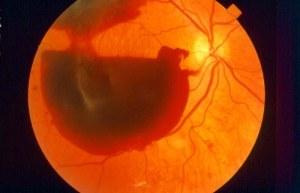
An epiretinal membrane (ERM), also known as a macular pucker, is a thin layer of scar-like tissue that forms on the surface of the macula—the central area of the retina responsible for sharp, detailed vision.
What is an epiretinal membrane?
An epiretinal membrane (ERM), also known as a macular pucker, is a thin layer of scar-like tissue that forms on the surface of the macula—the central area of the retina responsible for sharp, detailed vision.
Over time, this membrane may contract and create traction on the retina, causing the normally smooth surface of the macula to become wrinkled or distorted.
Symptoms of Epiretinal Membrane
Symptoms vary depending on the severity of the membrane and may affect one or both eyes.
Common symptoms include:
Common symptoms include:
- Blurred central vision
- Distorted vision (metamorphopsia), where straight lines may appear wavy
- Difficulty reading or recognizing fine details
- Changes in image size or shape compared with the other eye
- Gradual reduction in visual clarity
In mild cases, vision often remains stable and patients may continue their daily activities without significant limitations.
In more advanced cases, distortion and reduced vision may interfere with reading, driving, or other visually demanding tasks.
In more advanced cases, distortion and reduced vision may interfere with reading, driving, or other visually demanding tasks.
Causes of Epiretinal Membrane
Most epiretinal membranes develop as part of the natural aging process of the eye and are related to changes in the vitreous—the gel that fills the inside of the eye.
Possible causes include:
- Posterior vitreous detachment (the most common association)
- Previous retinal tears or retinal detachment
- Eye inflammation
- Eye trauma
- Retinal vascular diseases
- Previous eye surgery
During or after vitreous separation, cells may grow on the surface of the macula and form a thin membrane. As this membrane contracts, it can wrinkle the macular surface, similar to the way a thin piece of fabric folds when pulled.
Diagnosis of Epiretinal Membrane
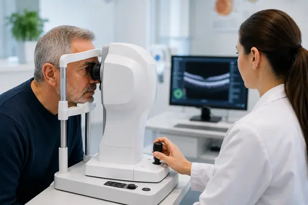
Diagnosis is made through a complete ophthalmic examination and specialized imaging tests.
-
Dilated retinal examination
Allows direct evaluation of the macula and detection of retinal surface changes.
-
OCT (Optical Coherence Tomography)
OCT is essential both for diagnosis and for monitoring progression over time.
-
Fluorescein angiography
Used in selected cases when additional information about retinal circulation or associated conditions is required.
OCT is crucial both for the initial diagnosis and for monitoring the condition over time.
Treatment of Epiretinal Membrane
Treatment depends mainly on the patient’s symptoms and the degree to which daily activities are affected.
Importantly, not every epiretinal membrane requires surgery.
-
Observation (early or mild cases)
For early or mild membranes:
- no medication is required
- eye drops and laser treatment are not effective
regular monitoring is usually sufficient
Many patients remain stable for years without requiring intervention.
- The vitreous humor that causes traction is removed
- The epiretinal membrane is carefully removed using specialized microsurgical instruments
- The procedure is performed on an outpatient basis
- At Athens Vision, we use sutureless techniques (minimally invasive transconjunctival vitrectomy)
After surgery, vision improves gradually and may exceed pre-membrane levels, depending on the initial damage and the tissue’s response.
Frequently Asked Questions
- Most of the vitreous, which exerts a pulling force on the macula, is removed
- A special gas is injected into the eye to act as an “internal tamponade” so that the hole closes
- The patient remains in a prone position (on their stomach) for 5–7 days so that the gas exerts constant pressure on the area
- The gas is gradually absorbed and replaced by fluid produced by the eye itself
At Athens Vision, we use modern, minimally invasive sutureless vitrectomy techniques whenever it is safe to do so.
Complications are rare, but they include:
- Infection
- Bleeding
- Retinal tear or detachment
- Reappearance of the membrane
- Accelerated onset of cataracts (typically expected with advancing age)
The ophthalmologist will discuss all the factors in detail to determine the best, personalized treatment plan.
A doctor who specializes in this condition

ALEXANDER CHARONIS
Ophthalmic Surgeon
Director of the Retina Department at AthensVision, Board Certified by the American and European Boards of Ophthalmology
Specialties: Vitreoretinal Surgery, Ocular Infections