Retinal detachment
The retina is the thin layer of nerve tissue located at the back of the eye. It receives light and converts visual information into signals that are transmitted to the brain.
It functions much like the film or sensor of a camera: if it is not properly positioned, the image we perceive becomes blurred or distorted.
What is the retina?
The retina is the thin layer of nerve tissue located at the back of the eye. It receives light and converts visual information into signals that are transmitted to the brain.
It functions much like the film or sensor of a camera: if it is not properly positioned, the image we perceive becomes blurred or distorted.
What is retinal detachment?
Retinal detachment is a serious eye condition in which the retina separates from its normal position at the back of the eye.
It is considered an ophthalmic emergency.
Causes of Retinal Detachment
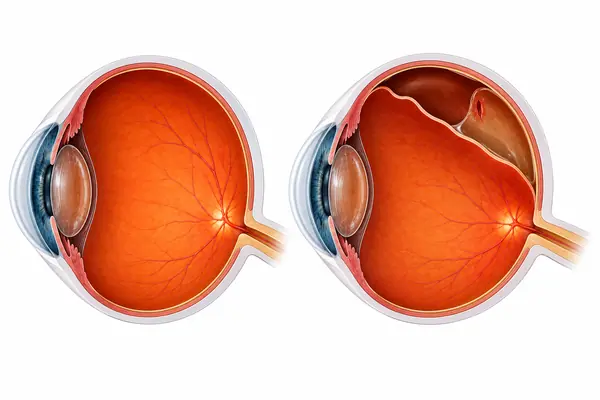
With age, the vitreous gradually shrinks and separates from the retina, a normal process called posterior vitreous detachment. In some cases, this separation creates traction on the retina and causes a retinal tear. Fluid can then pass through the tear and accumulate underneath the retina, causing it to detach—similar to wallpaper peeling away from a wall.
- High myopia (nearsightedness)
- Previous cataract surgery
- Glaucoma
- Previous eye injury
- Retinal detachment in the other eye
- Family history of retinal detachment
- Areas of retinal thinning or degeneration identified during retinal examination
Warning Signs of Retinal Detachment
Typical warning signs include:
- Flashes of light
- A sudden increase in floaters (especially when accompanied by flashes)
- A shadow or “black curtain” appearing in the peripheral vision
- A sensation that part of the visual field is missing or becoming dark

Diagnosis of Retinal Detachment
After the pupil is dilated with special eye drops, the retina can be carefully examined to identify retinal tears, areas of weakness, or an existing detachment.
In some patients, retinal tears or areas at increased risk for detachment may be detected during a routine eye examination before symptoms occur.
Treatment of a Retinal Tear
Treatment is performed with laser photocoagulation or cryotherapy, creating a protective scar around the tear that seals the retina and prevents fluid from passing underneath.
These procedures are usually performed in the office and cause minimal discomfort.
Treatment of Retinal Detachment
The choice of surgical technique depends on the type, location, and severity of the detachment.
Scleral Buckle Surgery
The procedure is usually performed under local anesthesia and does not require prolonged hospitalization.
Pneumatic Retinopexy
A specific head position is required for several days after the procedure. The gas bubble is gradually absorbed by the eye.
Vitrectomy
When gas is used, it gradually disappears and is naturally replaced by the eye’s own fluid.
In selected cases, vitrectomy may be combined with scleral buckle surgery.
Frequently Asked Questions
After the procedure, it is normal to experience mild discomfort. Flashes of light and floaters may persist for months. If gas was used, a specific head position is required for a period of time, and flying is prohibited until the gas is fully absorbed, as sudden changes in altitude can cause a dangerous increase in intraocular pressure. After recovery, a change in eyeglass prescription is often necessary.
All surgical procedures carry risks; however, a retinal detachment that is not treated promptly usually leads to severe and permanent vision loss. Possible complications include infection, bleeding, increased intraocular pressure, and the development of cataracts.
Although most surgeries are successful, in some cases a second surgery is required.
Vision recovery after a retinal detachment is gradual and can take months. In some patients, vision does not fully return, especially when the retinal detachment was extensive or had persisted for a long time before treatment.
With the help of modern technology, detachment can be successfully treated in 80–90% of cases with a single surgery; this rate rises to approximately 95% with additional procedures when necessary.
A doctor who specializes in this condition