

Glaucoma is a heterogenic group of disorders that damage the optic nerve and cause irreversible visual loss. Glaucoma is a chronic progressive optic neuropathy that is usually associated with elevated intraocular pressure. However, almost 1/3 of Caucasians have glaucoma without elevated intraocular pressure.
What causes increased intraocular pressure?
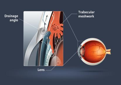
Intraocular pressure refers to the pressure of the fluid, called aqueous humor, which fills the front part of the eye. Aqueous humor is produced by a structure behind the iris called ciliary body. Aqueous moves continuously through the pupil, towards the angle of the eye formed by the iris and the cornea (the transparent dome covering the front part of the eye), and subsequently leaves the eye. A disruption in the balance between aqueous humor production and outflow causes increased intraocular pressure. It is also important to understand that eye pressure fluctuates throughout the day even in normal individuals.
Is intraocular pressure always increased in glaucoma?
As already discussed, elevated intraocular pressure is the most common and the only modifiable risk factor in glaucoma. That means that some optic nerves may be more susceptible to elevated intraocular pressure, whereas others may be more resistant to damage. About 1/3 of Caucasian patients develop “normal or low-tension glaucoma” where optic nerve damage occurs without elevated eye pressures. In this type of glaucoma mostly vascular factors such as diastolic arterial hypotension, sleep apnea or migraine headaches, may play a more important role. Therefore, increased intraocular pressure is not synonymous to “glaucoma”. Elevated intraocular pressure alone without optic nerve pathology is called ocular hypertension.
Normal Vision |
Early Glaucoma |
Advanced Glaucoma |
Near End-Stage Glaucoma |
Are there several types of glaucoma?
There are several types of glaucoma:
Primary Open Angle Glaucoma, Primary Angle Closure Glaucoma, Secondary Open Angle (i.e. Exfoliative Glaucoma), Secondary Angle Closure Glaucoma and Congenital Glaucoma represent some of them. These types do not carry a similar prognosis for the patients with some of them being associated with more severe disease.
Primary Open Angle Glaucoma
(Chronic Simple Glaucoma)
This is the most common type of glaucoma. The intraocular pressure increases slowly without any symptoms in the early stages of disease. For this reason, approximately 1/2 patients remain undiagnosed until late stages of the disease.
Primary Angle Closure Glaucoma
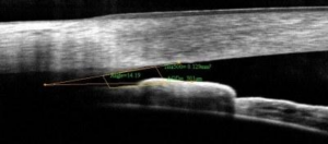
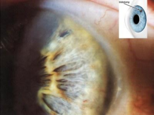
This type of glaucoma is more common in Asians and in people with hyperopia (farsightedness). The intraocular pressure may increase acutely (acute angle closure glaucoma) causing severe eye pain accompanied by colored halos, blurred vision, headache and nausea. Acute Angle Closure Glaucoma represents an ocular emergency and requires immediate attention. Alternatively, the rise in intraocular pressure may be more chronic and indolent in nature (Chronic Angle Closure Glaucoma). Common denominator in angle closure glaucoma is the iris obstructing the drainage pathway of the eye. This obstruction is usually relieved by a short laser procedure called peripheral iridotomy.
Secondary Glaucoma
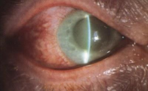
It can be either secondary open angle or secondary angle closure glaucoma. By definition in secondary glaucomas there are ocular or systemic factors that affect the intraocular pressure. Exfoliative or pseudoexfoliative glaucoma is by far the most common type of secondary open angle in Greece.
Congenital Glaucoma
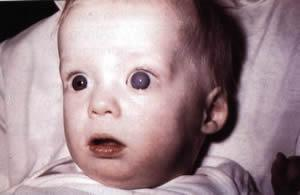
Congenital Glaucoma is a rare disease that occurs with a frequency of approximately 1:10.000 births. Boys are more commonly affected with 75% of cases being bilateral. It manifests with tearing, photophobia (light sensitivity) and enlargement of the eye (buphthalmos), which most commonly the caregiver notice. In the vast majority it requires surgical treatment. Some types of congenital glaucoma are associated with additional and more complex ocular pathology.
What are the risk factors for glaucoma?
1) Age
The prevalence of Primary Open Angle Glaucoma increases with age. It is uncommon under the age of 40 and the prevalence increases to about 2% after the age of 40 and to about 4% in people over the age of 80 years.
2) Race People of African descent are 3-4 times more likely to suffer from Primary Open Angle Glaucoma compared to Caucasians. Furthermore, in people of African descent the disease appears at a younger age and follows a more aggressive course. Asians on the contrary have an increased risk of suffering from angle closure glaucoma or low-tension glaucoma (glaucoma without elevated eye pressures).
3) Family History People with a family history of glaucoma (1st. degree relative) have a 3-4 higher likelihood to develop glaucoma themselves in their lifetime. Therefore, the American Academy of Ophthalmology recommends annual screening after the age of 40 for people with a family history of glaucoma, since the disease is asymptomatic in early stages.
4) Refractive Problems People with myopia may also have an increased risk of developing Primary Open Angle Glaucoma, whereas people with high degrees of hyperopia are predisposed to angle closure glaucoma.
5) Cardiovascular/Systemic Factors Patients with migraine headaches, Raynaud phenomenon, hypotension, sleep apnea syndrome are at increased risk of suffering from glaucoma (particularly of low-tension glaucoma). Normal or Low-Tension Glaucoma is a form of the disease, where intraocular pressure never exceeds normal limits.
6) Thin Cornea The cornea is the transparent dome of the eye located in front of the iris. A cornea that is thinner than average constitutes an independent risk factor for the development of glaucoma.
7) Diabetes Mellitus Patients with Diabetes may have an increased risk of developing glaucoma even though it is not clear if a causal relationship exists.
What should I do if I have an increased risk for glaucoma?
Individuals with risk factors for glaucoma can undergo a comprehensive eye examination including tonometry (measurement of the intraocular pressure) and dilated fundus examination (evaluation of the optic nerve). The American Academy of Ophthalmology recommends annual screening for individuals with 1st. degree relatives with glaucoma. If there are suspicious signs of glaucoma patients can undergo further testing including standard automated perimetry (visual field testing) and digital structural tests of the Optic Nerve Head or the Retinal Nerve Fiber Layer such as Optical Coherence Tomography (OCT), Scanning Laser Polarimetry (GDx) or Confocal Scanning Laser Ophthalmoscopy (HRT).


Can glaucoma be treated?
Damage caused by glaucoma to the optic nerve is irreversible but further progression can and must be prevented. Therefore, timely diagnosis and treatment is of paramount importance. Ultimate goal according to the European Glaucoma Society guidelines is to maintain visual function and quality of life. Unfortunately, when patients become symptomatic, noticing for example difficulties in adjusting to extreme or changing lighting conditions, considerable time has been wasted and significant optic nerve reserves have been lost.
What can treatment achieve?
Treatment reduces intraocular pressure in an effort to halt progression of the disease. Physicians consider a combination of factors to come up with a target intraocular pressure (IOP) needed to arrest progression individually. This “target IOP” needs to be maintained in order to inhibit further progression and usually is well below the so-called upper normal limit of 21mmHg. It is adjusted according the stage of the disease, life expectancy and the pressure level thought to be responsible for the damage that has already occurred.
What treatments are available for glaucoma?
Traditionally, the initial treatment for glaucoma consists of eye-drops aiming at reducing intraocular pressure. There are now six common families of such medications and your doctor will select the most suitable for you. While some drugs reduce the production of aqueous humor, others increase the outflow and some work both ways. Alternatively, a short, safe and painless laser procedure (selective laser trabeculoplasty) can be employed that lowers intraocular pressure alleviating the need for eyedrops in the majority of selected patients. For the drugs to be effective, they have to be used regularly and on a continuous basis. In selected patients, medical therapy is inadequate and surgical treatment is required.
How should I instill my eye-drops?
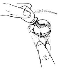
There are many effective ways to put the drops in. A simple way is to tilt your head backwards, to pull down the lower lid and drop the medicine into the space created between the eye and the eyelid. Subsequently, you should close your eyes and gently exercise pressure with your index finger at the inner corner of the eye for about 2-3 minutes in order to maximize absorption and to minimize systemic side effects. This way, the medication remains longer in contact with the eye. If you have been instructed to use more than one eye-drop at a given time you should space them at least 5 minutes apart.

Can glaucoma drops have side effects?
Like all medications, glaucoma eye-drops can cause local or systemic side effects. If you notice any side effect notify your doctor. Do not forget to tell your doctor about other health problems you may have or medications you are taking for other conditions.
The most common side effects include:
-Foreign body sensation -Red eye -Blurred vision -Itching -Allergic reactions -Contact dermatitis -Low heart rate or blood pressure -Difficulty breathing in predisposed individuals
Is a laser treatment suitable for glaucoma?
Laser treatments are available for certain categories of glaucoma. SLT (Selective Laser Trabeculoplasty) can lower the intraocular pressure by about 20% in selected patients with open angle glaucoma or ocular hypertension. Additionally, a different laser (YAG laser) can alter the natural course of angle closure glaucoma favorably if performed in a timely fashion and also prevent a crisis of acute angle closure glaucoma. Other options include Argon laser Iridoplasty and Diode laser Transscleral Cyclophotocoagulation. At the Athens Vision Eye Institute, experts in glaucoma perform all types of glaucoma laser surgery with state-of-the-art equipment.
When is surgery indicated for the treatment of glaucoma?
If medications or laser treatment fail to control intraocular pressure or if there is intolerance or non-compliance to medical treatment and the disease is progressing, we recommend surgery.
What types of glaucoma surgery do exist?
The most frequently employed first-line surgical procedure for glaucoma is called trabeculectomy with or without the ExPress device. In this procedure we create an alternative drainage pathway and divert the aqueous humor out of the eye, through a draining fistula into the sub-conjunctival space under the upper lid.
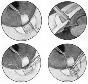
Other glaucoma surgical procedures include: Non-penetrating procedures such as deep sclerectomy or canaloplasty Ab-interno trabeculectomy, the Kahook Dual Blade procedure and i-Stent implantation Implantation of glaucoma drainage devices such as the Ahmed or the Baerveldt implant and cyclo-destructive procedures
Is surgery always successful?
Scientific evidence indicates that majority of patients will achieve low intraocular pressures (12-15mmHg) after a surgical procedure. The success rate of surgical treatment depends in general on the specific diagnosis (type of glaucoma), the selected procedure, race and age of the patient and on the presence of previous surgical interventions. In low-risk patients the success rate of trabeculectomy (the most commonly performed initial procedure) is well over 90% at the one-year mark. Postoperatively about 2/3 of patients will achieve satisfactory pressure control without medications (complete success). About 1/3-1/4 of patients will require topical medications (eye-drops) to sustain low intraocular pressures (qualified success) and a smaller proportion will require further surgery. Complications of surgical treatment include cataract formation, hypotony (too low intraocular pressure), infection (1:2000 cases) and severe bleeding (1:10000 cases).
How can we prevent visual loss due to glaucoma?
The visual loss in glaucoma can be prevented with timely diagnosis and treatment. Nevertheless, even in developed western societies about half of the glaucoma patients are undiagnosed as symptoms appear only in late stages of the disease. Experts stress that the quality of life of patients can suffer greatly as people with glaucoma demonstrate reduced ability to perform daily activities such as identifying objects, reading, driving, going up and down the stairs and recognizing faces.
Therefore:
If you are 40 years of age or older and have a family history of glaucoma you should undergo a comprehensive eye examination every year.
What is neuroprotection?
The concept of neuroprotection emerged in the late nineties. The idea is to make the neurons that constitute the optic nerve resistant to the process of programmed cell death called apoptosis which operates as glaucoma progresses. If, somehow, we can suspend the mechanism of apoptosis, neurons will survive irrespective to intraocular pressure. Our physicians have participated in key basic and clinical research trials that aimed at developing neuroprotective strategies and have also received worldwide recognition for their contribution. Unfortunately, there is no medication that has been approved for human use yet.
Currently, medications such as tacrolimus are tested at Harvard Medical School while at Johns Hopkins genetically engineered viruses are injected into the eye of study animals with the goal of changing the genetic environment of ganglion cells in order to make them more resistant to injury.
Glaucoma Summarized:
- Glaucomas are a group of disorders that cause optic nerve injury usually in association with increased intraocular pressure.
- Glaucoma is one of the most common eye diseases of the elderly. Approximately 2% of the population over 40 will develop some type of glaucoma with the incidence further increasing with age.
- It is estimated that currently in the countries of the European Union 9.25 million people suffer from glaucoma, a number that will approach 12.4 million in the next decade.
- If left untreated, glaucoma can cause blindness.
- Glaucomatous visual loss can be prevented if diagnosed early and treated appropriately.
Dr. Theodoros Filippopoulos, MD
Ophthalmic Surgeon