

SERVICES / RETINA
Retina and Macula
What the retina is, the main retinal symptoms, and how retinal conditions are diagnosed.
What Is the Retina?
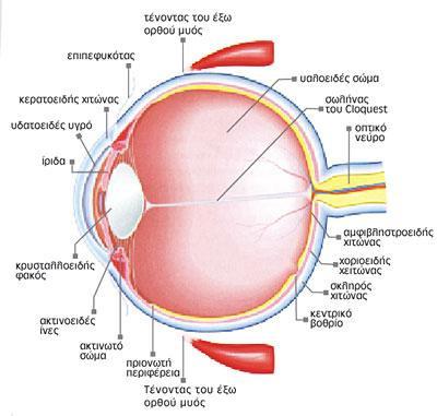
The retina is a light-sensitive layer of tissue that lines the inside of the eye. It receives light and sends the image to the brain. The eye works like a camera: the lens at the front of the eye focuses light onto the retina. You can think of the retina as the film in the camera.
Retina Department – Athens Vision
At the Retina Department of Athens Vision, we treat all retinal conditions using modern diagnostic and treatment methods, always with an emphasis on personalized care.
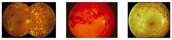
The Diabetic Retinopathy Unit provides complete monitoring and treatment, from digital angiography and OCT to laser, intravitreal drug therapy (injections) and minimally invasive vitrectomy. The Age-Related Macular Degeneration Unit offers early diagnosis, advanced treatments and low-vision support to maximize vision.
The Surgical Unit manages all surgical conditions, from retinal detachments and macular holes to complex reconstructive cases, using modern microsurgical techniques. The Pediatric Unit focuses on retinal diseases in children, particularly retinopathy of prematurity, with specialized diagnostic and treatment methods.
All units are supported by modern fundus imaging equipment and by collaboration with international institutes, for the application of new technologies and for second opinions in complex cases.
Retinal Symptoms
Flashes and Floaters
What are flashes and floaters?
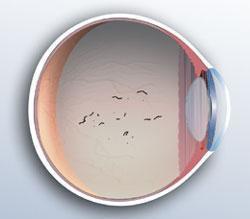
The inside of the eye contains the vitreous, a clear gel. Opacities within it create the familiar “floaters”: small spots or lines that are seen mainly against bright backgrounds (e.g., the sky or a white wall). They are often caused by normal age-related changes, high myopia or old hemorrhages, and usually become less noticeable over time.
When should I be concerned?
- If new floaters or flashes appear suddenly.
- If the number of floaters increases suddenly, with or without flashes.
In that case, an urgent visit to an ophthalmologist for a dilated retinal examination (fundoscopy) is needed, to rule out a retinal tear or retinal detachment. Flashes indicate mechanical traction on the retina by the vitreous, while new floaters may indicate the presence of blood cells. Both situations require immediate evaluation.
Metamorphopsia (Distortion of Central Vision)
Metamorphopsia is distortion of central vision, often noticed when reading or looking at straight objects. It is caused by problems in the macula, the area of the retina responsible for sharp vision and for recognizing fine detail.
Main causes:
- Age-related macular degeneration (particularly the wet form)
- Epiretinal membrane
- Macular hole, or macular edema (e.g., due to diabetes or retinal vein occlusion)
- Central serous chorioretinopathy
Diagnosis:
Metamorphopsia usually affects only one eye. It can be detected with simple tests: covering one eye and looking at straight lines (e.g., with the Amsler grid). Reading glasses should be worn for these tests.
What should you do?
If you notice distortions, see an ophthalmologist promptly for a dilated retinal examination. The doctor may request further tests, such as fluorescein angiography or optical coherence tomography (OCT), to assess the condition of the macula.
Diagnosis
Fundoscopy (Dilated Retinal Examination)
Fundoscopy is the examination of the “fundus” of the eye (the retina and vitreous) in order to diagnose disease.
How is it done, and is it painful?
- It requires mydriasis (dilation of the pupil) with eye drops, which take about 10–15 minutes to work.
- The doctor uses lenses together with a slit lamp or an indirect ophthalmoscope for a detailed examination.
- The procedure is not painful, only mildly uncomfortable.
- It takes 2–3 minutes, and a special lens or gentle pressure may be used to view the far periphery of the retina.
After the examination:
- Vision remains blurred for several hours (usually around 4–6), especially up close.
- Avoid driving until your vision has returned to normal.
- Sunglasses are helpful because of light sensitivity (photophobia).
Pregnancy and breastfeeding:
Fundoscopy is safe, and in some cases necessary, for the early diagnosis of conditions that threaten vision.
A doctor who specializes in this condition

Alexandros Charonis
Ophthalmic Surgeon
Director of the Retina Department, Athens Vision; Board Certified by the American and European Boards of Ophthalmology
Specialties: Vitreoretinal Surgery, Ocular Inflammation