

SERVICES / MACULAR DEGENERATION
Macular hole
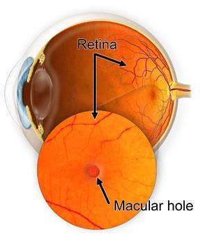
A macular hole is a tear or opening in the central part of the retina—the macula.
What is a macular hole?
A macular hole is a small opening in the central part of the retina—the macula—which is responsible for sharp, detailed vision.
When a macular hole develops, central vision becomes blurry or distorted, while peripheral vision usually remains normal. The macula is the “heart” of detailed vision, so even small changes can significantly affect daily activities such as reading or driving.
How does a macular hole form?
The interior of the eye is filled with the vitreous, a transparent gel-like substance. With age, the vitreous gradually liquefies and separates from the retina (posterior vitreous detachment).
When the vitreous remains strongly attached to the macula, this separation may create traction forces that pull on the retina and lead to the formation of a macular hole.
Other factors that may contribute include:
-
Other factors that may contribute include:
- Chronic macular edema
- Previous macular conditions that weaken the retinal tissue
Symptoms of a macular hole
In the early stages, a macular hole causes gradual changes in central vision.
The most common symptoms include:
The most common symptoms include:
- Blurred central vision
- Distortion of straight lines (metamorphopsia)
- Difficulty reading fine print
- In advanced stages: development of a central blind spot (scotoma), affecting both near and distance vision
Despite the central vision loss, peripheral vision almost always remains intact.
Diagnosis of a macular hole
Diagnosis is made by an ophthalmologist using:
-
Dilated retinal examination:
A detailed examination of the retina to directly identify changes in the macula.
-
OCT (Optical Coherence Tomography):
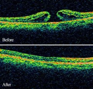
High-resolution imaging of the macula that accurately shows the size, depth, and characteristics of the hole.
At Athens Vision, we use state-of-the-art spectral-domain OCT technology, allowing precise visualization and monitoring of macular diseases.
Treatment of a macular hole
The most effective treatment is vitrectomy, a highly specialized microsurgical procedure.
Frequently Asked Questions
- Most of the vitreous, which exerts a pulling force on the macula, is removed
- A special gas is injected into the eye to act as an “internal tamponade” so that the hole closes
- The patient remains in a prone position (on their stomach) for 5–7 days so that the gas exerts constant pressure on the area
- The gas is gradually absorbed and replaced by fluid produced by the eye itself
At Athens Vision, we use modern, minimally invasive sutureless vitrectomy techniques whenever it is safe to do so.
Vision gradually improves after the hole closes, but rarely returns fully to pre-injury levels.
The final outcome depends on:ω
- the size of the hole
- the duration of the damage
- the tissue’s individual response
Like any surgical procedure, vitrectomy carries potential complications, which are, however, rare when performed by experienced surgeons:
- Infection
- Bleeding
- Retinal detachment
- Increase in intraocular pressure
- Cataracts (common in older adults)
Your ophthalmologist will discuss the benefits and potential risks in detail so that the most appropriate treatment approach can be determined.
A doctor who specializes in this condition

ALEXANDER CHARONIS
Ophthalmic Surgeon
Director of the Retina Department at AthensVision, Board Certified by the American and European Boards of Ophthalmology
Specialties: Vitreoretinal Surgery, Ocular Infections